

TREATMENT DETAILS
Panoramic
A radiographic examination that presents the upper and lower jaw, teeth, and bone structure in a single 2D digital image to guide diagnosis and treatment planning.
OVERVIEW
Comprehensive diagnosis and safe planning.
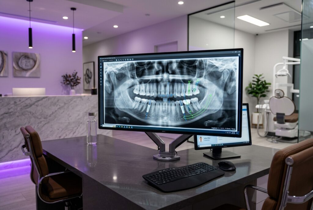
A panoramic radiograph is a 2D digital dental X-ray showing the upper and lower jaws and all teeth on these jaws in a single plane.
Thanks to dental panoramic films, which assist in making an accurate and comprehensive diagnosis during the initial examination, many pre-treatment preparations—such as interproximal caries undetected by visual examination, bone defects, bone loss levels, wisdom teeth positions, implant planning, treatment planning, and prosthesis planning—are completed thoroughly[cite: 15]. Caution should be exercised when taking panoramic films during the early stages of pregnancy and in the first year following radiotherapy[cite: 15]. However, with technological advancements in digital panoramic film devices used in recent years, the radiation level administered has decreased significantly[cite: 15]. In short, panoramic films are among our indispensables in the initial examination[cite: 15]. In cases where this film is insufficient, periapical or bitewing radiographs can be taken[cite: 15].
01
DIGITAL EVALUATION
02
CUSTOM PLANNING
EVALUATION
How is suitability determined?
Treatment selection is made by evaluating clinical examination, necessary imaging, and personal expectations together.
01
Clinical necessity
Imaging needs are determined by evaluating current complaints, overall oral health, suspected caries, and bone condition.
02
Systemic status check
Pregnancy (especially the first trimester) and recent history of radiotherapy are checked; protective measures are planned.
03
Radiographic method selection
The overview of a panoramic film is evaluated against the necessity of regional detail radiographs such as periapical or bitewing.
“A panoramic radiograph illuminates deep tissues invisible to the clinical eye, forming the foundation for accurate diagnosis and safe planning.”
Dr. Ediz Cem Türk
TREATMENT PROCESS
Step-by-step process
01
Initial preparation
Earrings, necklaces, hairpins, glasses, and removable dentures (if any) are removed; a protective lead apron is fitted.
02
Positioning
The head and chin are positioned at the correct symmetry and angle on the device’s bite block using laser guide lights.
03
Fast and comfortable exposure
The unit quietly rotates around the head in about 12-15 seconds to complete the digital panoramic scan.
04
Digital analysis and planning
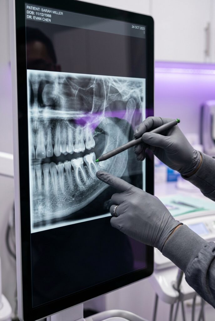
The high-resolution image is transferred to the screen in seconds; a treatment plan is created together with the patient.
FREQUENTLY ASKED
Detailed answers
It is a radiographic method that presents the lower and upper jaws, teeth, and bone structure in a single 2D digital image to guide diagnosis and treatment planning.
Panoramic radiography displays the jawbones, root structures of teeth, temporomandibular joints, and sinus cavities on a single wide-angle digital film. It is one of the essential diagnostic methods indispensable during the initial examination. It enables early detection of interproximal caries, bone defects, bone loss levels, wisdom teeth positions, root apex lesions, and cysts. Additionally, it provides the practitioner with a comprehensive anatomical map prior to implant surgery, prosthetic frameworks, orthodontic treatment, and general treatment planning.
It can be evaluated in adults and children visiting for an initial examination, undergoing general oral screening, or requiring surgical, implant, and orthodontic planning.
It is widely preferred in new patient examinations to thoroughly determine current oral and jaw status. It serves as an important initial step for individuals with impacted or unerupted teeth, temporomandibular joint (TMJ) disorders, suspected widespread caries or bone loss, and those starting implant or orthodontic treatment. In children, it is also safely used to monitor the development and eruption direction of permanent tooth buds.
Metallic items and dentures are removed, pregnancy and medical history are inquired about, and a protective lead apron is fitted.
Earrings, necklaces, piercings, hairpins, glasses, and removable dental prostheses that could affect image quality and create shadows (artifacts) are removed prior to the procedure. The patient’s systemic health status is inquired about; specifically, suspected pregnancy or recent history of radiotherapy is checked. As a standard safety protocol, the patient is fitted with a lead apron that blocks radiation scatter.
The patient is positioned at the device’s bite block, and the unit rotates around the head in about 12-15 seconds to transfer the digital image to the screen.
The patient stands in front of the device and gently bites on a bite block protected by a disposable sterile sheath using their front teeth. Laser guide lights position the head in correct symmetry, supported by head holders. During exposure, the device rotates around the patient’s head with a smooth motion for approximately 12-15 seconds. The patient remains motionless with their tongue pressed against the palate. Data collected by digital sensors is instantly projected onto the computer screen in high resolution.
It is a completely painless, non-invasive, and contact-free procedure; as no film is placed inside the mouth, it does not trigger a gag reflex.
Panoramic radiography is an extraoral (outside the mouth) technique. Because no rigid film plate is placed inside the mouth, it is extremely comfortable for patients experiencing gagging or nausea. No mechanical pressure, pain, or stinging is felt during exposure. It is sufficient to stand motionless for a few seconds while the unit rotates around the head.
Preparation and exposure take approximately 1-2 minutes in total; the digital image is ready on-screen within seconds.
Removing metal accessories and positioning correctly in the unit takes 1 minute, while the device rotation around the head takes about 12 to 15 seconds. Thanks to digital technology, there is no processing or chemical development time; the image is instantly delivered in high resolution to the practitioner’s workstation screen and evaluated together with the patient in the same session.
Unless essential, it is postponed during the first trimester of pregnancy and within the first year post-radiotherapy, or applied following physician consultation.
Because the early stages of pregnancy (first 3 months) represent organogenesis, X-rays are not recommended unless there is an urgent and essential clinical presentation, or are postponed until after childbirth. Caution should also be exercised during the first year for patients receiving radiotherapy, deciding through oncologist consultation and special protective measures in imperative situations. It can be applied safely to all children and adults capable of standing upright or sitting upright in a wheelchair in the device.
For higher resolution in a specific tooth or bone area, periapical, bitewing, or 3D CT scans can be taken.
Panoramic films provide a general two-dimensional overview. However, for millimeter details of a tooth’s root apex lesion or clarity on initial interproximal caries, it can be supported with periapical or bitewing radiographs. In complex surgical and implant cases where bone thickness must be examined in depth, 3D dental volumetric tomography (DVT/CBCT) can be included in the evaluation.
With new-generation digital sensors, radiation levels are significantly lower compared to conventional films, equivalent to daily environmental background radiation.
Thanks to advancing digital imaging technology, the amount of X-rays in modern panoramic radiography units has been reduced to minimal levels. The average radiation dose received in a digital panoramic exposure is similar to natural background radiation exposed in nature over a few days or radiation received during a 1-2 hour airplane flight. This minimal risk is minimized further through protective lead apron usage.
It is used as a standard diagnostic step during the initial examination; the entire treatment plan is created by sharing it on the digital screen with the patient.
In our clinic, panoramic radiography is used actively during the initial examination as the starting point for accurate and predictable treatment. The exposure taken is instantly transferred to the practitioner’s screen; the condition of teeth, bone levels, potential cysts, and angles of 20-year-old teeth (wisdom teeth) are examined in detail on-screen together with the patient. Implant, prosthesis, surgical, or restorative treatments are planned individually in light of this objective data.
EXAMINATION AND PLANNING
Let’s evaluate the right option for you together.
The suitability of Panoramic Radiograph application for you can only be determined after clinical evaluation.